Home
Uncategories
Anatomy Rib Cage Muscles : The Intercostal Muscles Intercostal Spaces Youtube / They are further categorized according function such as flexion, extension, or rotation.
Anatomy Rib Cage Muscles : The Intercostal Muscles Intercostal Spaces Youtube / They are further categorized according function such as flexion, extension, or rotation.
Anatomy Rib Cage Muscles : The Intercostal Muscles Intercostal Spaces Youtube / They are further categorized according function such as flexion, extension, or rotation.. Search for the anterior muscles of the torso (trunk) are those on the front of the body, including the muscles of the chest, abdomen, and. Rib anatomy landmarks lungs and ribs anatomy rib anatomy numbers 10th rib anatomy floating ribs anatomy thorax surface anatomy 1st rib anatomy lower rib anatomy human anatomy rib cage muscles rib cage structure typical rib anatomy single rib anatomy anterior. Rib cage pain can be caused. The last time i had these was last friday night, and they lasted for two hours. In the muscular system, muscle tissue is categorized into three distinct types:
This is a table of skeletal muscles of the human anatomy. It encloses and protects the heart and lungs. The muscles that affect the knee's movement run along the thigh and calf. The ribs also provide attachment sites for thoracic muscles. Rib cage pain may be sharp, dull, or achy and felt at or below the chest or above the navel on either side.
Two Minutes Of Anatomy Ribcage Youtube from i.ytimg.com It forms the bony framework for breathing. Surface anatomy of the brainstem. The transversus thoracic muscles originate from the posterior surface of the xiphoid process and the lower part of the body of the sternum. Anatomy rib cage muscles / human ribs diagramnumbered. The subcostal muscles are found in the inferior portion of the thoracic wall. The upper edge is round and the lower sharp. Our latest youtube film is ready to run. Rib cage mechanics and muscles.
The thoracic cage is a component of the thoracic wall and encloses the majority of the structures of the respiratory system.
Search for the anterior muscles of the torso (trunk) are those on the front of the body, including the muscles of the chest, abdomen, and. The direction of the fibres parallels that of the innermost intercostal. In spite of its resistance, the cage is dynamic, allowing pulmonary ventilation to. The thoracic cage is a component of the thoracic wall and encloses the majority of the structures of the respiratory system. The thoracic cage consists of the 12 thoracic vertebrae, the associated intervertebral discs, 12 pairs of ribs with their costal cartilages, and the sternum. #proko #art #anatomy #ribs #ribcage #humananatomy #tutorial In this rib bones anatomy quiz, you can test your knowledge of the ribs. The last time i had these was last friday night, and they lasted for two hours. Rib cage pain may be sharp, dull, or achy and felt at or below the chest or above the navel on either side. Play games, take quizzes, print and more with easy notecards. Muscle spasms felt within the rib cage may also be caused by the abdominal muscles. It forms the bony framework for breathing. You'll need a bench and one dumbbell to do this exercise.
The last time i had these was last friday night, and they lasted for two hours. Rib cage mechanics and muscles. The last two, the floating ribs, have their cartilages ending in the muscle in the abdominal wall. The serratus anterior is a muscle that attaches your shoulder blade, known as your scapula, to your rib cage. It forms the bony framework for breathing.
Airway Anatomy And Physiology Clinical Essentials Paramedic Care Part 2 from what-when-how.com The superior or upper border of each rib, at the costal groove 9 insertion: This muscle assists in depression of the ribs. The thoracic cage is a component of the thoracic wall and encloses the majority of the structures of the respiratory system. Human muscles · april 17, 2020. The major abdominal muscles include the transverse abdominals, the rectus abdominis, and the external and internal oblique muscles. It impairs full expansion of the ribcage, thus affecting the oxygen content of the blood. Tendons attach the muscles to each other. This is a table of skeletal muscles of the human anatomy.
This muscle assists in depression of the ribs.
The space between each rib is called the intercostal space, and there are 11 intercostal spaces in the thoracic cage, which are filled with nerves, lymph nodes, arteries, veins, and muscles.in fact, when you eat ribs at a restaurant, you're eating the intercostal muscles of an animal. The serratus anterior is a muscle that attaches your shoulder blade, known as your scapula, to your rib cage. Muscle spasms felt within the rib cage may also be caused by the abdominal muscles. Rib cage mechanics and muscles. Anatomy of the rib cage diagram. The rib cage has three important functions: It impairs full expansion of the ribcage, thus affecting the oxygen content of the blood. The thoracic cage consists of the 12 thoracic vertebrae, the associated intervertebral discs, 12 pairs of ribs with their costal cartilages, and the sternum. The dome shaped thoracic cage provides the necessary rigidity for organ protection, weight support for the upper limbs and anchorage for muscles. They also contract involuntarily, but have a. The ribs are a set of twelve paired bones which form the protective 'cage' of the thorax. The inferior or lower border of the rib above 9 the fibers of the internal intercostals lie perpendicular to those of the. The major abdominal muscles include the transverse abdominals, the rectus abdominis, and the external and internal oblique muscles.
#proko #art #anatomy #ribs #ribcage #humananatomy #tutorial It forms the bony framework for breathing. The subcostal muscles are found in the inferior portion of the thoracic wall. Our latest youtube film is ready to run. In most undergraduate anatomy courses, you will need to understand the key landmarks on a typical rib bone, as well as general information about the ribs and the thoracic cage.
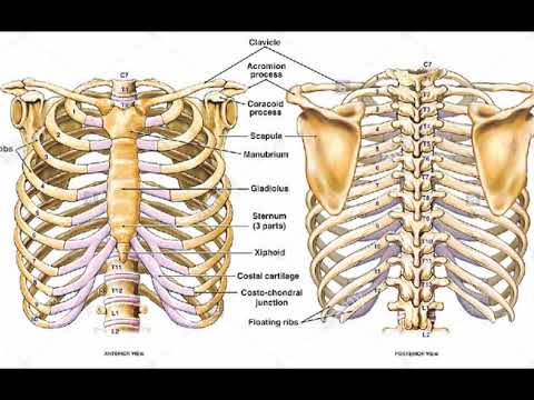
Surgical Anatomy Of The Chest Wall Thoracic Key from i0.wp.com With the upper ribs, closer to the nodule (and in the case of lower ribs, a little further from the nodule) they are curved and have a rough surface that connects them with muscles, angulus costae. Anatomy the rib cage is a bony structure found in the chest (thoracic cavity). In this image, you will find thoracic vertebrum, costochondral joint, costal cartilage, costal margin, costal arch, thoracic vertebrum, xiphoid process, xiphisternal joint, body, manubrial sternal joint, manubrium, the sternal notch in it. The muscles that affect the knee's movement run along the thigh and calf. Click the image to watch the anatomy of the rib cage video. They articulate with the vertebral column posteriorly, and terminate anteriorly as cartilage (known as costal cartilage). The upper edge is round and the lower sharp. Muscle spasms felt within the rib cage may also be caused by the abdominal muscles.
Human muscles · april 17, 2020.
The transversus thoracic muscles originate from the posterior surface of the xiphoid process and the lower part of the body of the sternum. Our latest youtube film is ready to run. They comprise of thin slips of muscle, which run from the internal surface of one rib, to second and third ribs below. Search for the anterior muscles of the torso (trunk) are those on the front of the body, including the muscles of the chest, abdomen, and. With the upper ribs, closer to the nodule (and in the case of lower ribs, a little further from the nodule) they are curved and have a rough surface that connects them with muscles, angulus costae. Tendons attach the muscles to each other. The inferior or lower border of the rib above 9 the fibers of the internal intercostals lie perpendicular to those of the. If you know where muscles attach and how. Human rib cage anatomy model. The primary responsibilities of the ribcage involve protecting the thoracic visceral organs, enclosing the thoracic visceral organs, and is included in the. Play games, take quizzes, print and more with easy notecards. It encloses and protects the heart and lungs. The fibres pass superolaterally to insert into the internal surface of costal cartilages of ribs two to six.
They are further categorized according function such as flexion, extension, or rotation anatomy rib cage. The rib cage intrinsically holds the muscles of respiration (diaphragm, intercostal muscles, etc.) that are crucial for active inhalation and forced exhalation, and therefore has a major ventilatory function in the respiratory system.


0 Comments:
Post a Comment