Home
Uncategories
Arteries Diagram Easy / arteries of heart with diagram - Clip Art Library - Arteries are blood vessels that carry blood away from the heart to other tissues.
Arteries Diagram Easy / arteries of heart with diagram - Clip Art Library - Arteries are blood vessels that carry blood away from the heart to other tissues.
Arteries Diagram Easy / arteries of heart with diagram - Clip Art Library - Arteries are blood vessels that carry blood away from the heart to other tissues.. Arteries should always be depicted using a red color; Each artery is a muscular tube lined by smooth tissue and has three layers: Blood supply to the scalp. How to make a circulatory system diagram Labels include cephalic vein, brachial artery/vein, basilic vein, musculoskeletal nerve, ulnar collateral artery, radial collateral artery, ulnar nerve/artery/vein, interosseous artery/vein, median nerve and radial nerve/artery/vein.
This is done to make it easy to distinguish between arteries and veins as they look almost identical. Diagram of blood vessel types each time your heart beats, blood is forced into large arteries. The media, a layer of muscle that lets arteries. Arteries of the cardiovascular system diagram practice test. Plaque in arteries is caused by deposits of ldl, which is commonly known as bad cholesterol.
Circulatory System & Heart | gcse-revision, pe-physical-education, anatomy-and-physiology ... from revisionworld.com Origin right aortic sinus (lower origin than lca) course down right av groove toward crux of the heart, gives off pda (85%) from which septals arise, continues in lav groove giving off posterior lv branches (posterolaterals). We are pleased to provide you with the picture named blood circulation principal veins and arteries diagram.we hope this picture blood circulation principal veins and arteries diagram can help you study and research. Anatomynote.com found blood circulation principal veins and arteries diagram from plenty of. For more anatomy content please follow us and visit our website: Plaque in arteries is caused by deposits of ldl, which is commonly known as bad cholesterol. The gonadal arteries are paired arteries that send blood to the testes in males and the ovaries in females. The media, a layer of muscle that lets arteries. It originates from the heart and branches out into smaller arteries which supply blood to the head region (brachiocephalic artery), the heart itself (coronary arteries), and the lower regions of the body.
Although there are different types of circulatory system diagrams, you fill find some consistencies throughout.
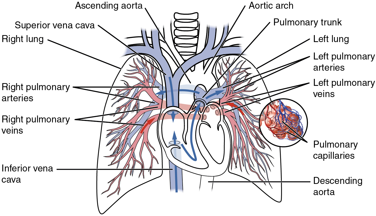
The right ventricle receives blood from the right atrium and pumps it to the lungs, where it is loaded with oxygen. The gonadal arteries are paired arteries that send blood to the testes in males and the ovaries in females. Diagram of blood vessel types each time your heart beats, blood is forced into large arteries. A major artery in the neck, one vessel it supply's is the basilar artery, and it basically suppl's the entire brain with blood. Arteries are blood vessels that carry blood away from the heart to other tissues. We are pleased to provide you with the picture named blood circulation principal veins and arteries diagram.we hope this picture blood circulation principal veins and arteries diagram can help you study and research. The aorta is the main systemic artery and the largest artery of the body. Each artery is a muscular tube lined by smooth tissue and has three layers: It originates from the heart and branches out into smaller arteries which supply blood to the head region (brachiocephalic artery), the heart itself (coronary arteries), and the lower regions of the body. Left anterior descending artery (lad) the left coronary arteries supply: Origin right aortic sinus (lower origin than lca) course down right av groove toward crux of the heart, gives off pda (85%) from which septals arise, continues in lav groove giving off posterior lv branches (posterolaterals). Finally, the smallest arteries, called arterioles are further branched into small capillaries, where the exchange of all the nutrients, gases and other waste molecules are carried out. Veins are the blood vessels present throughout the body.
Arteries are the blood vessels that carry blood away from the heart, where it branches into even smaller vessels. The right ventricle receives blood from the right atrium and pumps it to the lungs, where it is loaded with oxygen. Arteries of the cardiovascular system diagram practice test. Arteries should always be depicted using a red color; Plaque in arteries is caused by deposits of ldl, which is commonly known as bad cholesterol.
This artery transports de-oxygenated blood away from the ... from cdn.thinglink.me Arteriosclerosis occurs when the blood vessels that carry oxygen and nutrients from your heart to the rest of your body (arteries) become thick and stiff — sometimes restricting blood flow to your organs and tissues. We are pleased to provide you with the picture named blood circulation principal veins and arteries diagram.we hope this picture blood circulation principal veins and arteries diagram can help you study and research. The left atrium receives oxygenated blood from the lungs and pumps it to the. The vertebral arteries, and the internal carotid arteries. Anatomy of the nerves, arteries and veins of the arm (upper extremity). Your email will not be shown with your comment. This vessel supports the anterior circle of. Origin right aortic sinus (lower origin than lca) course down right av groove toward crux of the heart, gives off pda (85%) from which septals arise, continues in lav groove giving off posterior lv branches (posterolaterals).
The basilar artery is part of the vertebrobasilar system and is one the the major arteries in the brain.
We are pleased to provide you with the picture named blood circulation principal veins and arteries diagram.we hope this picture blood circulation principal veins and arteries diagram can help you study and research. This is done to make it easy to distinguish between arteries and veins as they look almost identical. For more anatomy content please follow us and visit our website: The media, a layer of muscle that lets arteries. The aorta is the main systemic artery and the largest artery of the body. Finally, the smallest arteries, called arterioles are further branched into small capillaries, where the exchange of all the nutrients, gases and other waste molecules are carried out. The heart is a muscular organ with four chambers. This branch of the abdominal aorta divides into the internal and external. Origin right aortic sinus (lower origin than lca) course down right av groove toward crux of the heart, gives off pda (85%) from which septals arise, continues in lav groove giving off posterior lv branches (posterolaterals). Your email will not be shown with your comment. The intima, the inner layer lined by a smooth tissue called endothelium. The tunica medica, which is the very muscular middle layer in arteries, is thinner and less muscular in veins. Veins should be depicted using blue.
Spend a few minutes analysing the diagram, and trying to connect the location of the structures with what you've learned in the video. Diagram of the cardiac anatomy showing the right and left side of the heart. A very good labled with diagram easy to study. How to make a circulatory system diagram Veins are the blood vessels present throughout the body.
Circulatory Pathways · Anatomy and Physiology from philschatz.com This branch of the abdominal aorta divides into the internal and external. A very good diagram, made the study of coronary arteries of anterior heart wall very easy. The gonadal arteries are paired arteries that send blood to the testes in males and the ovaries in females. The heart can be divided into 2 sides, a right and a left. Simple heart diagram arteries and veins heart arteries biology drawing science diagrams physical education lessons a level biology critical care nursing rn nurse The walls of the arteries are tightly and closely bound to the. The heart is a muscular organ with four chambers. While it can't be completely eliminated or undone, you can manage it and reduce the risk of a blockage.
Arteries are the blood vessels that carry blood away from the heart, where it branches into even smaller vessels.
The basilar artery is part of the vertebrobasilar system and is one the the major arteries in the brain. Labels include cephalic vein, brachial artery/vein, basilic vein, musculoskeletal nerve, ulnar collateral artery, radial collateral artery, ulnar nerve/artery/vein, interosseous artery/vein, median nerve and radial nerve/artery/vein. Artery, in human physiology, any of the vessels that, with one exception, carry oxygenated blood and nourishment from the heart to the tissues of the body. Spend a few minutes analysing the diagram, and trying to connect the location of the structures with what you've learned in the video. Blood supply to the scalp. This is done to make it easy to distinguish between arteries and veins as they look almost identical. Finally, the smallest arteries, called arterioles are further branched into small capillaries, where the exchange of all the nutrients, gases and other waste molecules are carried out. The media, a layer of muscle that lets arteries. The left atrium receives oxygenated blood from the lungs and pumps it to the. Your email will not be shown with your comment. The gonadal arteries are paired arteries that send blood to the testes in males and the ovaries in females. Arteries should always be depicted using a red color; We are pleased to provide you with the picture named blood circulation principal veins and arteries diagram.we hope this picture blood circulation principal veins and arteries diagram can help you study and research.
Arteries of the brain and 'circle of willis' diagram there is a point at which the anterior and posterior arterial circuits of the brain unite or anastomose arteries diagram. The intima, the inner layer lined by a smooth tissue called endothelium.


0 Comments:
Post a Comment